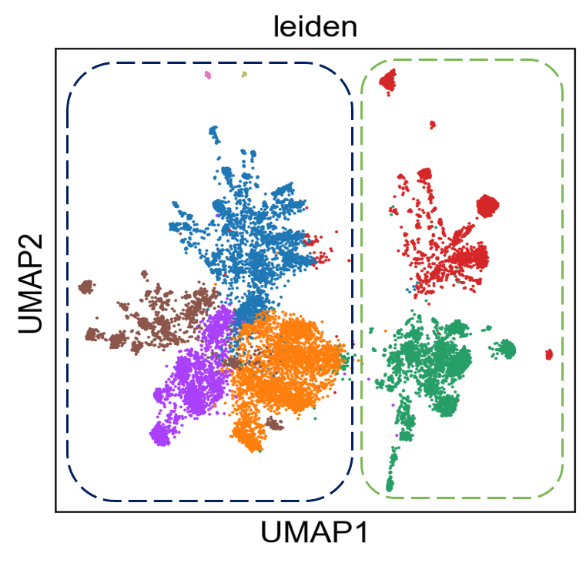
Reveal subtle cell states
The resolving power to distinguish precise transcriptional states
Traditional cell biology and microscopy recently merged with genomics to create spatial omics, a powerful new approach for studying biology. A range of methods and techniques are emerging, most of which target one class of biomolecules, often compromising sensitivity, specificity or dynamic range in favor of higher plex.
Rebus Biosystems has developed the Rebus Esper™, an integrated platform that combines resolution-enhancing Synthetic Aperture Optics (SAO) with automated fluidics to enable multiple types of assays for detecting biomarkers at single-molecule, subcellular resolution. The streamlined workflow minimizes hands-on time and reduces variability by automating the laborious steps required for multiplexed single-molecule detection. Several assay chemistries will be available for the Rebus Esper platform.
The Esper High Fidelity Assay for spatial transcriptomics automates single-molecule FISH to quantify 30 custom genes across ~1 cm2 of tissue in ~2.5 days. Upcoming assays will combine dual detection of protein staining with quantification of RNA expression, increase plex levels to hundreds of genes, and enable the use of FFPE tissue, a challenging but important sample type for clinical and translational research. A flexible and open data analysis pipeline is integral to the Rebus Esper platform.
Data can be processed and viewed using the provided tools, which can be integrated with publicly available or commercial analysis packages. With advanced optics, automated fluidics, and intuitive software, the Rebus Esper makes spatial omics accessible to any biologists seeking to visualize the molecular complexity of life.
The Esper High Fidelity Assay for spatial transcriptomics automates single-molecule FISH to quantify 30 custom genes across ~1 cm² of tissue in ~2.5 days.